
fMRI Explained: What BOLD Signals Really Measure (And Brain Imaging's Hidden Limits)
Imagine staring at a vibrant brain scan, splashed across a magazine cover: glowing hotspots pulsing like city lights at night, promising to unlock the secrets of love, fear, or even free will. It's mesmerizing. But what if that rainbow spectacle isn't capturing thoughts zipping between neurons? What if it's something far more mundane—a vascular blush? Welcome to the real world of fMRI explained, where the BOLD signal steals the show, and brain imaging limits remind us that neuroscience methods are powerful yet profoundly indirect.
Functional magnetic resonance imaging, or fMRI, has revolutionized how we peek inside the living brain. Yet beneath the hype lies a method rooted in blood flow, not direct neural fireworks. Let's demystify it, step by step, for those hungry for the evidence behind the headlines.
The Heart of fMRI: Decoding the BOLD Signal
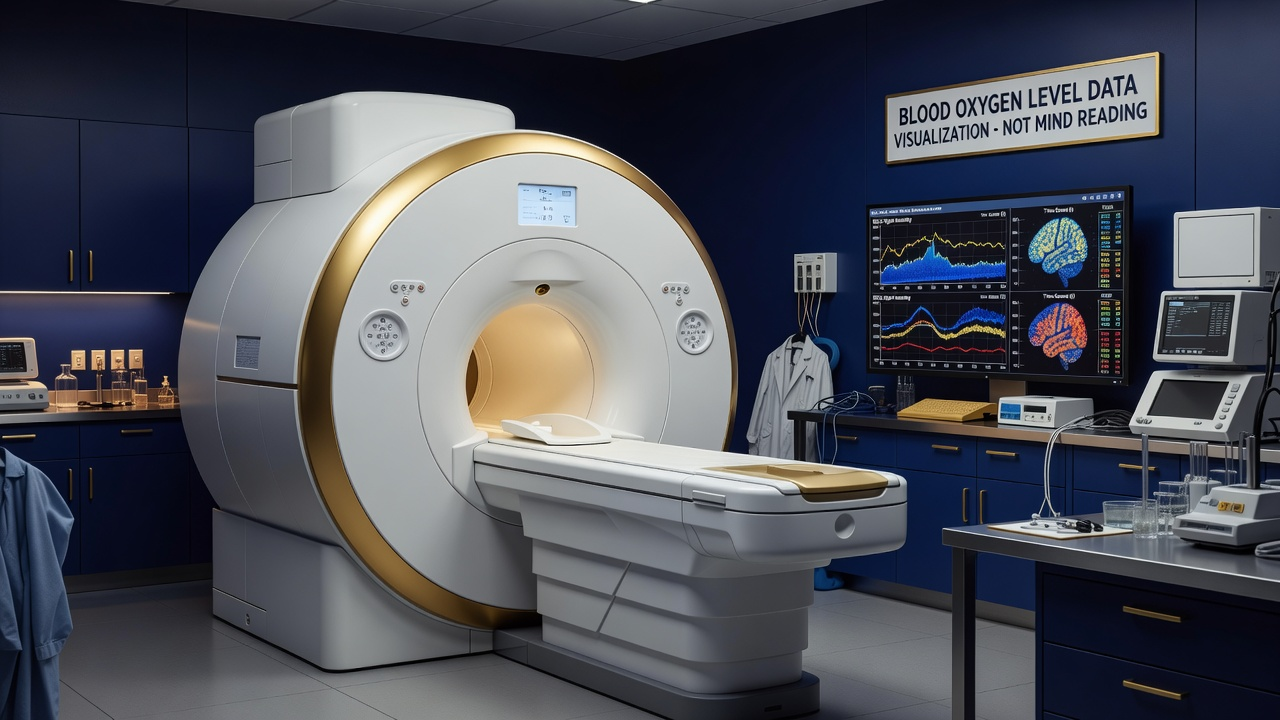
At its core, fMRI doesn't eavesdrop on neurons firing. Instead, it tracks the BOLD signal—Blood-Oxygen-Level-Dependent. When a brain region ramps up activity, neurons guzzle oxygen. Local blood vessels respond by flooding the area with oxygen-rich blood, more than the neurons need. This overcompensation creates a magnetic signature detectable by MRI scanners.
Picture it: a bustling neural neighborhood calls for reinforcements. Arteries dilate, hemoglobin levels shift, and the scanner lights up that zone in red or yellow. It's a proxy, elegant but delayed—peaking seconds after the actual neural event. This vascular lag is the first clue in understanding brain imaging limits: fMRI maps metabolic demand, not thoughts or decisions in real time.
From Raw Data to Brain Maps: The Art of Subtraction and Contrasts
Experimental Design in Action
fMRI experiments hinge on clever design. Subjects might view faces versus objects, or rest versus a task. Researchers subtract baseline scans from task scans, hunting signal differences. These contrasts yield activation maps, but only where changes exceed noise.
Enter statistical thresholding: raw data drowns in variability—head motion, breathing, scanner drift. Software applies tests like t-tests, setting p-value cutoffs (often 0.001) and correcting for multiple comparisons across thousands of voxels. A cluster of suprathreshold voxels emerges as "activated." It's rigorous neuroscience methods, yet one volunteer's fidget can skew results.
- Subtraction assumes clean baselines—no hidden carryover effects.
- Contrasts isolate conditions, but real cognition blends seamlessly.
- Thresholding balances sensitivity and false positives, often visualized as colorful blobs.
The Reverse Inference Trap: Why a Lit-Up Blob Isn't "The Love Center"
Here's where headlines crumble. A hotspot in the ventral striatum during romantic gaze? Tempting to declare it "the seat of love." But that's reverse inference: from brain activity to mental state, without direct proof. The region activates for reward, pain, money—context reigns.
Activity shows correlation, not causation. A brain area lights up with love, but does it cause love? Rarely proven.
Poldrack's seminal warning: without validating tasks, claims falter. Localization seduces—"the" prefrontal cortex for decision-making—but brains are distributed, plastic networks.
Unveiling Brain Imaging Limits: Motion, Replication, and Beyond
Practical Pitfalls
Motion artifacts top the list: a millimeter head shift rivals BOLD changes. Sophisticated corrections help, but not perfectly. Low signal-to-noise demands large samples, yet early studies skimped, fueling replication woes.
Spatial resolution hovers at 2-3mm—coarser than cortical columns. Temporal blur from hemodynamics misses millisecond dynamics. Individual variability? Massive. Group averages mask personal brains.
- Motion corrupts: even "still" subjects drift.
- Replication crisis: many findings evaporate in meta-analyses.
- Indirect measure: BOLD tracks ensembles, not single cells.
Bridging to Psychology and AI: Bounded Comparisons
These brain imaging limits echo in psychology, where fMRI informs but doesn't dictate behavior models. Explore our Psychology Door for behavioral evidence unburdened by scanner confounds.
In AI, fMRI inspires neural nets, yet bounded analogies prevail—no BOLD in silicon. Check the Artificial Intelligence Door for grounded parallels at Aetheria AI.
The fMRI Journal: What It Reveals—and Hides
fMRI offers stunning snapshots of brain metabolism, pinpointing where oxygen surges during tasks. It excels in group-level patterns, validating circuits from animal models. Yet it whispers, never shouts: no direct neural code, no causal arrows without intervention.
Next time a scan dazzles, ask: What's the BOLD telling us? A vascular vote of confidence in neural hustle, filtered through stats and design. In neuroscience's grand quest, fMRI is a vital tool—flawed, indirect, indispensable. Approach with awe, and skepticism.