
Neuroscience Methods Demystified: EEG, fMRI, Single-Unit Recording & Brain Imaging Limitations Explained
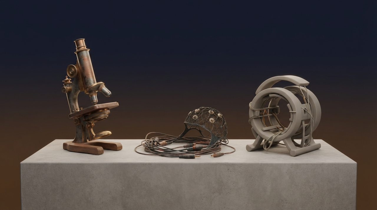
Picture this: a surgeon's scalpel slices through gray matter in the 19th century, revealing tangled fibers under a microscope. Or fast-forward to today, where a person lies still in a massive magnet, their thoughts lighting up colorful brain scans. Neuroscience methods promise windows into the mind, but what do they truly reveal? For intellectually curious explorers in Aetheria's Knowledge Library, this guide demystifies core techniques—from post-mortem anatomy to cutting-edge optogenetics. We'll unpack what each measures directly versus what it infers, their resolutions in time and space, inherent limits, and the dance between correlation and causation. Along the way, consider how these tools, much like instruments in science's grand observatory, shape the questions we ask about consciousness.
Foundational Pillars: Post-Mortem Anatomy and Lesion Studies
Neuroscience began humbly, with knives and microscopes. Post-mortem anatomy directly measures brain structure after death—tracing neuron clusters, fiber tracts, or lesions via staining techniques. Pioneers like Santiago Ramón y Cajal visualized individual cells, establishing neurons as the brain's basic units. Spatial resolution? Down to micrometers. Temporal? None; it's a static snapshot.
Lesion studies infer function from absence. Stroke or tumor damage disrupts abilities—like forgetting faces after temporal lobe injury—suggesting that region's role. Causal hints emerge: remove the part, lose the skill. Yet limits loom. Brains compensate via plasticity; one lesion rarely tells the whole story. Correlation (damage links to deficit) doesn't prove sole causation—networks overlap. Replication across patients builds confidence, but individual variability breeds uncertainty.
Electrophysiology: EEG and Single-Unit Recording in Neuroscience Methods
EEG: Capturing Brain Waves Non-Invasively
Electroencephalography (EEG) places electrodes on the scalp to record electrical potentials from millions of neurons firing in synchrony. It directly measures voltage fluctuations—brain waves like alpha rhythms during relaxation. Temporal resolution shines: milliseconds, ideal for tracking rapid events like seizure onset or attention shifts. Spatial? Poor—centimeters at best, smeared by skull and scalp.
Inferences abound: EEG correlates theta waves with memory encoding, but causation? Not directly. Artifacts from blinks or muscles muddy signals, demanding rigorous cleaning. Replication varies; protocols differ across labs.
Single-Unit Recording: Peering at Single Neurons
For precision, single-unit recording plunges microelectrodes into animal brains (or rarely, human surgery). It directly captures spikes from one neuron's action potentials—firing rates tied to stimuli, like a visual cell responding to edges. Temporal resolution: microseconds. Spatial: pinpoint, tens of micrometers.
Limits? Invasive, short-term, animal-centric. Infers coding schemes (rate vs. timing), but population dynamics escape single cells. Causation tests via stimulation exist, yet ethical bounds and variability challenge replication.
Imaging Frontiers: MRI, fMRI, PET, and Brain Imaging Limitations
Structural and Functional MRI
Magnetic Resonance Imaging (MRI) directly maps anatomy via proton alignments in magnetic fields—revealing gray matter volume or white matter integrity at millimeter resolution. Functional MRI (fMRI) infers activity from blood-oxygen-level-dependent (BOLD) signals: active neurons demand more oxygenated blood. Spatial: 1-3 mm. Temporal: seconds, lagging true neural events.
fMRI spotlights networks during tasks, like the default mode during mind-wandering. But BOLD correlates with metabolism, not direct electricity—veins, not neurons, drive signals. Causation? No; it shows engagement, not necessity.
PET: Tracing Metabolism
Positron Emission Tomography (PET) tracks radioactive tracers for glucose use or neurotransmitters. Spatial: 4-6 mm. Temporal: minutes. Infers hyperactivity in disorders like schizophrenia, but radiation limits use, and low resolution blurs details.
Across imaging, brain imaging limitations persist: measurement uncertainty from motion, noise, or indirect proxies. Spatial-temporal trade-offs mean no method nails both.
Optogenetics: Light-Controlled Precision in Animal Models
Optogenetics engineers neurons to fire (or silence) with light pulses via light-sensitive proteins. Directly causal: stimulate fear circuits in mice, induce panic. Spatial: cell-type specific. Temporal: milliseconds. Yet, it's animal-only, genetic tinkering alters baselines, and scaling to humans remains distant. Open questions: Does it mirror natural dynamics?
Navigating Uncertainty: Correlation, Causation, and Open Frontiers
All neuroscience methods grapple with inference. EEG and fMRI excel at correlation—activity patterns during thoughts—but causation demands intervention, like lesions or optogenetics. Replication crises highlight variability: small samples, p-hacking, diverse brains. Uncertainty fuels curiosity, echoing science's observational ethos and consciousness debates—do methods bias us toward localizable traits?
- Trade-offs define tools: EEG's speed vs. fMRI's detail.
- Multimodal fusion (EEG-fMRI) mitigates single-method flaws.
- Ethical replication prioritizes diverse cohorts.
Your Next Step: Journal the Mystery
These neuroscience methods illuminate the brain's vast terrain, yet shadows of uncertainty invite wonder. Pause: What personal experience— a vivid dream, a stubborn habit—might these tools probe? Journal it. Sketch a brain map of your day. In Aetheria's doors to science and consciousness, your reflections bridge method to meaning. The universe unfolds, one careful measure at a time.